tree in bud opacities treatment
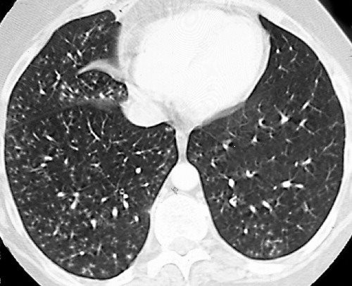
Normally they are. His chest CT images showed multi-focal patchy ground-glass opacities and parenchymal consolidation with both ill- and well-defined opacities predominantly involving the peripheral and posterior regions of both lungs.

Bronchial Cyst Azygoesophgeal Recess Typical Location Cysts Bronchial Radiology
The nodules occur when epithelial cells cover a necrotic area creating a necrobiotic nodule which is the cause of the cavity.

. Upper lobe parenchymal opacities with atelectasis secondary to bronchial obstruction are also commonly seen. Cavitating nodular opacities in the course of rheumatic diseases are much rarer than interstitial pulmonary pneumonias and vasculitides. Imaging can be further assessed with high-resolution CT scan which can also detect mucous plugging tree-in-bud opacities ground-glass attenuation and atelectasis.
These are most often located in the periphery or subpleurally. Appearances typical of COVID-19 Figure Figure7. In cases of early invasive aspergillosis CT scan can provide an early diagnosis.
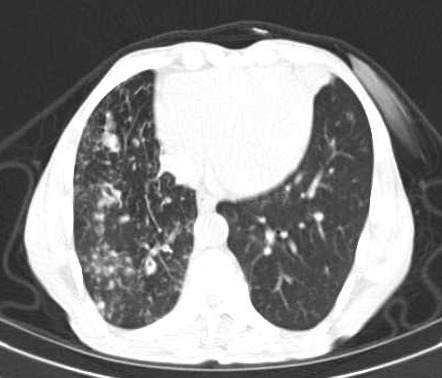
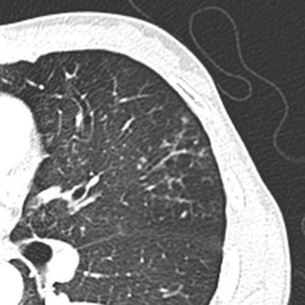
He unusually also had a focal area of tree-in-bud opacification in the right lower lobe. They may vary in size and wall thickness.
2

Tree In Bud Opacities With False Positive Gaffky Score And Diffuse Aspiration Bronchiolitis Mogi 2020 Journal Of General And Family Medicine Wiley Online Library

Pdf Tree In Bud Semantic Scholar
2
Tree In Bud Appearance Endobronchial Spreading Of Pulmonary Tuberculosis Radiology Case Radiopaedia Org
Tree In Bud Sign Lung Radiology Reference Article Radiopaedia Org

Pin On Tip

Tree In Bud Sign Lung Radiology Reference Article Radiopaedia Org

References In Causes And Imaging Patterns Of Tree In Bud Opacities Chest

Bronchiolitis Radiology Reference Article Radiopaedia Org Radiology Bud Pattern

Pin On Pulmonar
2

Pin Em Funny Animals

Chest Ct With Multifocal Tree In Bud Opacities Diffuse Bronchiectasis Download Scientific Diagram
View Of Tree In Bud The Southwest Respiratory And Critical Care Chronicles
Tree In Bud Sign An Overview Sciencedirect Topics
2
2
Tree In Bud Sign Lungs